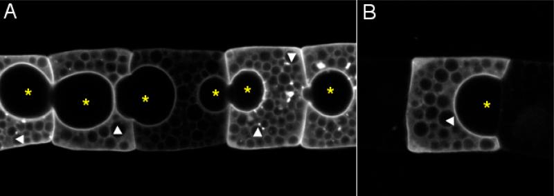
Figure 5.
Extracellular pockets in notochord column. A membrane-targeted GFP is expressed in notochord cells in mosaic pattern by electroporation. (A) A string of notochord cells expressing membrane-bound GFP at different levels. (B) An isolated GFP-positive cell. * indicates extracellular pockets. Arrowheads indicate intracellular yolk granules. Anterior to the left.

