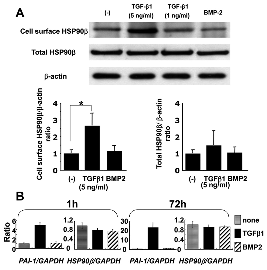
Figure 4. TGF-β1 regulates cell surface expression of HSP90β.
A, MG63 cells were starved and then incubated with either TGF-β1 (5 or 1 ng/ml) or BMP2 (50 ng/ml). 3 days after incubation, cell surface proteins were biotinylated and cell surface proteins and total proteins were collected and immunoblotted, as described in Figure 2. The amount of the proteins used for cell surface precipitant and total HSP90β were quantified by the amount of β-actin. Each value represents the mean of triplicate determinations; bars, mean + SD. *P < 0.05 B, MG63 cells were starved and then incubated with TGF-β1 (5 ng/ml) or BMP2 (50 ng/ml). Total RNAs were extracted from the cells at 1 h and 72 h after treatment. PAI-1 and HSP90β expression levels were analyzed by quantitative real-time PCR. Each value represents the mean of triplicate determinations; bars, SD.

