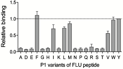
Fig. 4.
Peptide P1 anchor residue specificity of HLA-DR1 analyzed by yeast codisplay. Cells codisplaying HLA-DR1 and FLU peptides with the indicated P1 residue substitutions were analyzed by flow cytometry (Fig. S3) and normalized relative binding levels were calculated from fluorescence intensities (Materials and Methods). The dashed line represents binding to the wild-type FLU peptide with Tyr at P1. Error bars represent standard error of the mean determined from four independent experiments.

