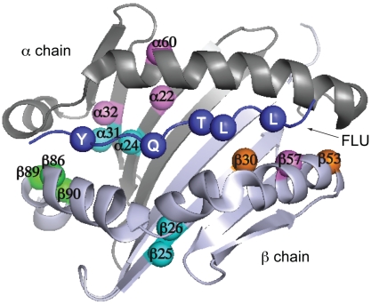
Fig. 6.
Mutation sites conferring altered P1 anchor specificity on HLA-DR1. Positions mutated in S4V1.5 (cyan), S4A1.9 (orange), S4E1.3 (magenta), or all three clones (green) are highlighted on a top view of the α1 and β1 domains, with the peptide anchor residue positions P1, P4, P6, P7, and P9 also highlighted (blue). Images generated from coordinates in PDB file 1DHL.

