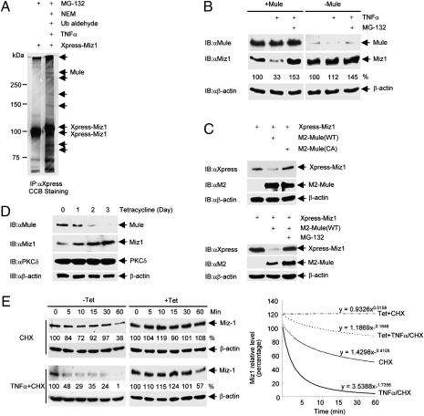
Fig. 1.
Mule regulates TNFα-induced Miz1 degradation. (A) HEK293 cells (1 × 10⁷) were transfected with expression vector encoding Xpress-Miz1 (50 μg). After 36 h, cells were pretreated with MG-132 for 2 h before stimulation with TNFα (5ng/mL) for 10 min. Cells extracts were harvested in Co-IP buffer supplemented with NEM and ubiquitin aldehyde. Miz1 was immunoprecipitated by anti-Xpress antibody, and the immune complexes were resolved on a 7.5% gel and visualized by CCB staining. Bands for mass spectrometry analysis to obtain tryptic peptides for the E3 ligase Mule were indicated by arrows. (B) WT MEFs transfected with control siRNA or siRNA for Mule (100 nM each) were treated with TNFα (5 ng/mL, 15 min) in the absence or presence of MG-132. Expression levels of Mule, Miz1 and β-actin were determined by immunoblotting. Miz1 protein levels were quantitated and normalized to β-actin levels, and the untreated controls were calculated as 100%. Similar results were obtained from at least three independent experiments. (C) HEK293 cells (1 × 10⁵) were transfected with Xpress-Miz1, along with M2-Mule (WT or the C4341A mutant) or empty vector (2 μg each), in the absence (Upper) or presence (Lower) of MG-132. Expression levels of Xpress-Miz1 and M2-Mule were determined. (D) U2OS stable cells harboring tet-inducible shMule (U2OS/shMule) were treated with tetracycline (2 μg/mL) for the indicated times. Expression levels of Mule, Miz1, PKCδ, and β-actin were determined. (E) U2OS/shMule stable cells were treated with or without tetracycline for 3 d followed by the addition of CHX (3 μg/mL) with or without TNFα (10 ng/mL) for the indicated times. Expression levels of Miz1 and β-actin were determined (Left). Miz1 protein levels were quantitated and normalized to β-actin levels, as shown in power trendlines (Right). Slope constants were indicated. Similar results were obtained from at least three independent experiments.

