Fig. 2.
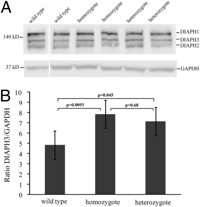
Expression of DIAPH3 protein is significantly increased in lysates from LCLs from heterozygotes and homozygotes vs. controls. (A) Immunoblot of LCL lysates from two wild types, two homozygotes, and two heterozygotes using DT154 antibody (43, 44). The DIAPH3 band is at 137 kDa (UniProt database); cross-reactivity is seen with DIAPH1 (140 kDa) and DIAPH2 (126 kDa). GAPDH was used as a loading control, and DIAPH3 densitometry was normalized to GAPDH densitometry. (B) Estimated marginal means and SE for the DIAPH3/GAPDH ratio for wild types, homozygotes, and heterozygotes tested in four batches of cells. There was a significant difference between controls and heterozygotes (4.83 vs.7.14, P = 0.045, a 1.48-fold increase) and between controls and homozygotes (4.83 vs. 7.85, P = 0.0093, a 1.62-fold increase). There was no significant difference in protein levels between homozygotes and heterozygotes (P = 0.68).

