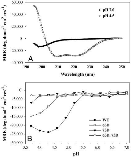
Fig. 2.
CD studies on the WT and mutant (57–98) HA2 peptides. (A) Far UV CD spectra of WT (57–98) HA2 peptide at pH 7.0 (•) and 4.5 (○), 25 °C, depicting the random coil to helix transition upon acidification. (B) pH titration of WT and mutant (57–98) HA2 peptides. The θ222 of the WT (•), 63D (○), 73 D (▾), and 63D, 73D (▵) mutant peptides are plotted as a function of pH. 73D mutant and 63D, 73D double mutant peptides are highly destabilized and do not form a coiled coil at any pH. Lines through the points are only for visual clarity.

