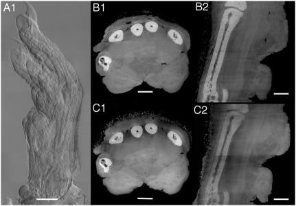
Fig. 4.
Imaging of a rat paw. (A) Differential phase-contrast radiography (7 stacks, RP protocol), (B1) axial and (B2) coronal slices through the paw acquired with the PS protocol, (C1) axial and (C2) coronal slices through the same sample obtained with the RP protocol. Structural details of both soft tissue (muscles, fat) and hard tissue (bone) are well visible. Scale bars, 2 mm (A), 1 mm (B1–2 and C1–2).

