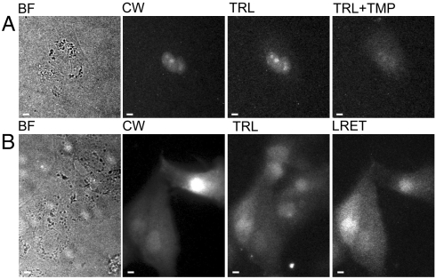
Fig. 3.
Both streptolysin O (SLO)-mediated membrane permeabilization and osmotic lysis of pinocytic vesicles deliver TMP-Lumi4 to the cytoplasm of MDCKII cells, and specific labeling of eDHFR fusion proteins can be visualized by time-resolved luminescence microscopy. (A), (B) Micrographs: BF, bright field; CW, continuous wave fluorescence (λex = 480 ± 20 nm, λem = 535 ± 25 nm); TRL, time-resolved luminescence (λex = 365 nm, λem > 400 nm, gate delay = 10 μs); TRL + TMP, 20 min after addition of TMP (final conc. = 10 μM) to culture medium; LRET, time-resolved luminescence (λex = 365 nm, λem = 520 ± 10 nm, gate delay = 10 μs). TRL and TRL + TMP images were adjusted to identical contrast levels. Complete details of time-resolved microscopy parameters are provided in SI Materials and Methods and Table S2. Scale bars, 10 μm. (A) MDCKII cells transiently cotransfected with DNA encoding nucleus-localized CFP and nucleus-localized eDHFR were incubated with TMP-Lumi4 (15 μM) and streptolysin O (SLO, 50 ng/mL) for 10 min. Time-resolved detection of broadband (> 400 nm) terbium emission reveals localization of TMP-Lumi4 in nucleus of transfected cell (corresponding to continuous wave fluorescence image of CFP emission). A time-resolved image taken 20 min. after addition of TMP (final conc. = 10 μM) to the culture medium shows diminished nuclear luminescence because TMP out-competes TMP-Lumi4 for binding to eDHFR. (B) MDCKII cells transiently transfected with DNA encoding GFP-eDHFR. Cells were incubated in hypertonic medium containing TMP-Lumi4 (50 μM) for 10 min to allow pinocytosis and subsequently exposed to hypotonic medium to effect lysis of pinocytic vesicles and release of probe into the cytoplasm. Time-resolved detection of broadband emission (> 400 nm) reveals terbium luminescence in TMP-Lumi4-loaded cells. Luminescence resonance energy transfer (LRET) is seen only in transfected cells (indicated in continuous wave fluorescence image) coincidentally loaded with TMP-Lumi4 when visualized in time-resolved mode through a narrow-pass filter (520 ± 10 nm), indicating specific labeling of the GFP-eDHFR fusion protein.

