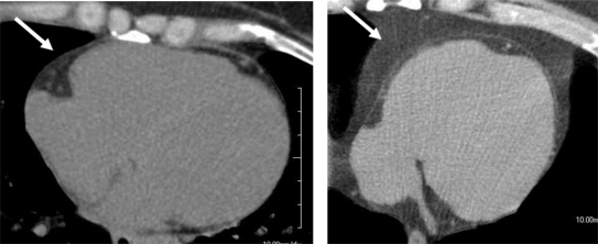
Figure 4.
A single, mid LV slice from a noncontrast CT of the heart (ie, a ‘heartscan’) demonstrating the epicardial fat distribution in a patient without metabolic syndrome (Left arrow) and a patient with known metabolic syndrome (right arrow). See text for details.
Abbreviations: LV, left ventricle; CT, computed tomography.

