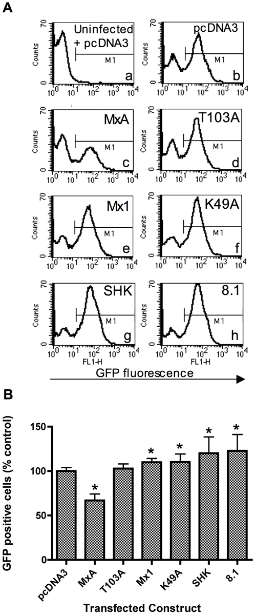
Figure 1. Chicken Mx proteins do not inhibit NDV-directed gene expression.
293T cells were co-transfected with a plasmid expressing the DsRed-express fluorescent protein and either pcDNA3 or a plasmid expressing the indicated Mx protein (human MxA, the MxA mutant T013A, murine Mx1, the Mx1 mutant K49A, wild type SHK (Asn631) or 8.1 (Ser631) chicken Mx). 48 h post-transfection, the cells were infected with NDV-GFP at an MOI which achieved approximately 60% infection (as determined by flow cytometry). 15 h post-infection, the cells were fixed and analysed by flow cytometry. Cells were then gated according to their expression of DsRed, and analysed for GFP fluorescence in the FL1-H channel. Panel A shows representative histograms for the GFP fluorescence in DsRed positive cells that were co-transfected with the indicated plasmids. Sub-panel (a) shows the background GFP fluorescence in uninfected, pcDNA3-transfected cells. The fluorescence threshold marker (M1) demarcates between GFP negative and positive cells. Panel B shows data derived from 6 replicates and bar heights show the % GFP positive cells expressed relative to that for the pcDNA3 control. The mean (and SD) are shown for DsRed positive cells co-transfected with the constructs as indicated. * indicates a significant difference (Students t-test) relative to pcDNA3 (p<0.05).

