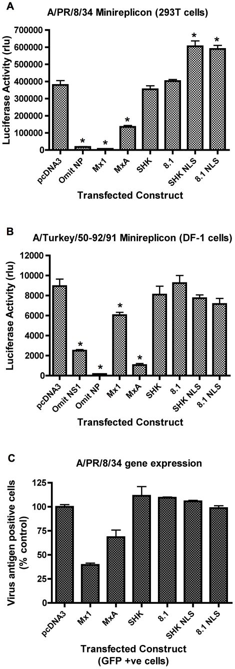
Figure 4. Nuclear localized chicken Mx lacks anti-influenza activity.
Panel A: Effect on influenza A/PR/8/34 minireplicon system. 293T cells were co-transfected with plasmids expressing the PB1, PB2, PA and NP proteins of influenza A/PR/8/34, a plasmid encoding a luciferase minireplicon, and a SEAP-expressing plasmid, together with either pcDNA3 or a plasmid expressing the indicated Mx protein (murine Mx1, human MxA, wild type chicken Mx proteins SHK (Asn631) and 8.1 (Ser631) and their nuclear-localised counterparts SHK NLS and 8.1 NLS). 48 h post-transfection, luciferase activity was measured and is shown as SEAP-corrected relative light units (rlu). The mean (and SD) of 3 replicates is shown. * indicates p<0.05 (Students t-test) relative to pcDNA3. Panel B: Effect on influenza A/Turkey/England/50-92/91 minireplicon system. DF-1 cells were transfected with plasmids encoding A/Turkey/England/50-92/91 PB1, PB2, PA and NP, A/PR/8/34 NS1, an influenza minireplicon plasmid encoding luciferase, and a SEAP expressing plasmid together with pcDNA3 or a plasmid expressing the indicated Mx protein (murine Mx1, human MxA, wild type chicken Mx proteins SHK (Asn631) and 8.1 (Ser631) and their nuclear-localised counterparts SHK NLS and 8.1 NLS). 48 h post-transfection, luciferase activity was measured and is shown as relative light units (rlu). The mean (and SD) of 6 replicates is shown. * indicates p<0.05 (Students t-test) relative to pcDNA3. Panel C: Effect on influenza A/PR/8/34 gene expression. 293T cells were co-transfected with Mx-expressing plasmids (or pcDNA3) and pEGFP-C1 and infected after 48 h with influenza A/PR/8/34. 15 h post-infection, the cells were stained for influenza vRNP and analysed by flow cytometry. The percentage of antigen positive cells is expressed relative to that of the pcDNA3 control. The mean (and range) of 2 replicates is shown for GFP positive cells co-transfected with the indicated constructs.

