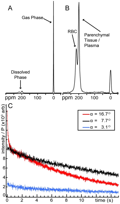
Figure 1. HP 129Xe MR signal intensity in human lungs.
(A) NMR spectrum obtained using a hard, 7° RF pulse. The gaseous HP 129Xe signal is used as a 0 ppm reference. (B) Spectrum from a selective, 7° pulse centered at 218 ppm. The 218-ppm peak arises from 129Xe dissolved in the red blood cells (RBC), and the 197-ppm arises from 129Xe in the blood plasma and semi-solid parenchymal tissues. (C) Dissolved HP 129Xe signal dynamics during single breath-hold radial imaging. Data points represent the magnitude of k-zero from each radial view weighted by the initial HP 129Xe polarization (P). Even using a relatively large flip angle of α∼17° and a rapid TR of 4.2 ms, substantial dissolved signal is still observed at the end of the breath-hold period due to rapid, diffusive replenishment of dissolved 129Xe magnetization.

