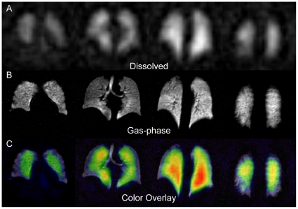
Figure 2. HP 129Xe MR imaging.
Panels are arranged with the more anterior portions of the lungs shown to the left and posterior portions to the right. (A) 15-mm-thick sections from a dissolved-phase HP 129Xe image (12.5×12.5 mm2 in-plane resolution) of a healthy human volunteer. (B) Corresponding 15-mm-thick slices from a gas-phase HP 129Xe image of the same subject (3.2×3.2 mm2 in-plane resolution). (C) Dissolved 129Xe image from (A) displayed in color and overlaid on the grayscale ventilation image from (B).

