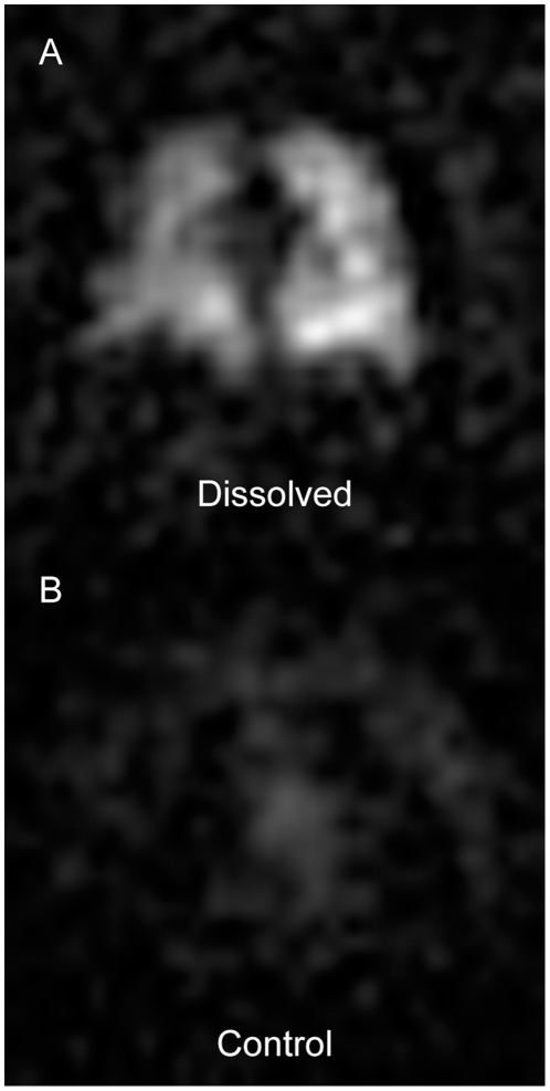
Figure 3. Off-resonant excitation of gas-phase 129Xe magnetization.
(A) Representative 15-mm-thick section from a standard dissolved HP 129Xe image (α = 8°, RF centered 3826 Hz above the gas-phase resonance) of a supine subject. (B) Corresponding 15-mm-thick section from a control image of the same supine subject. The MRI acquisition parameters were identical to those used to produce the image in (A) except that the RF pulse was centered 3826 Hz below the gas-phase resonance. Windowing and leveling were identical for both (A) and (B).

