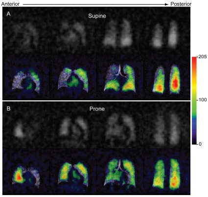
Figure 4. Postural dissolved HP 129Xe image heterogeneity.
Dissolved 129Xe images displayed in grayscale (top) and overlaid in color (bottom) on the corresponding ventilation images. For the color overlays, the dissolved image signal intensity (arbitrary units) is indicated in the legend. The ventilation image was obtained in the supine position. (A) Dissolved image acquired after the subject had been supine for 1 hour. Note, the more gravitationally dependent, posterior portions of the lungs exhibited higher signal intensities than did the less dependent anterior regions. (B) Same subject imaged 10 minutes after moving to the prone position. Again, the gravitationally dependent (now anterior) regions display increased signal intensity.

