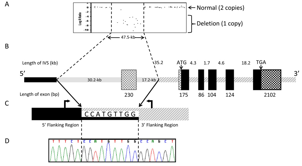
Fig. 2.
Experimental and schematic representations of homozygous deletion in Rab27A gene of patient with Griscelli Type 2 Syndrome.
(A) Illumina® 1M-Duo DNA Analysis BeadChip results illustrating a large scale deletion.
(B) Schematic basis of 47.5-kb deletion relative to the most abundant transcript of the Rab27A gene (GenBank NM_004580). The deletion (shaded gray line) removes the first exon, which is non-coding. The deletion does not contain any coding sequence. Cross-hatched boxes indicate 5’-UTR of the transcript and the checkered box indicates 3’-UTR of all transcripts. Black boxes indicate coding region of Rab27A.
(C) Two primers (block arrows) were used to amplify across the deletion that occurs within the 9-bp stretch shared by the two regions.
(D) Sequence analysis of the breakpoint region. Diagrams are not to scale.

