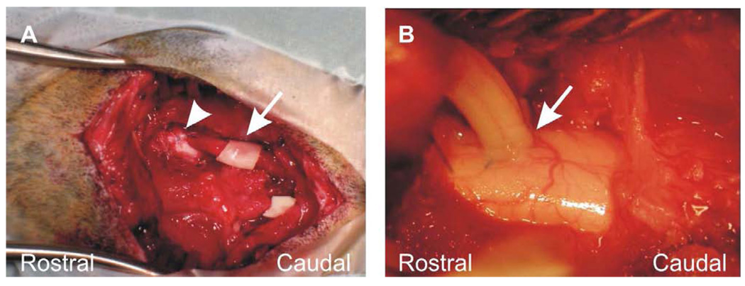
Figure 2.
Apposition of the peripheral nerve graft. (A) Rostral (arrowhead) and caudal ends are apposed to the spinal cord. Protective PVA/PVP film (arrow) was placed around the mid portion of the graft. The piece of PVA/PVP film at the lower right of the field is not associated with the graft. (B) Demonstration of the distal apposition (arrow) of the PNG to the right side of the upper thoracic cat spinal cord.

