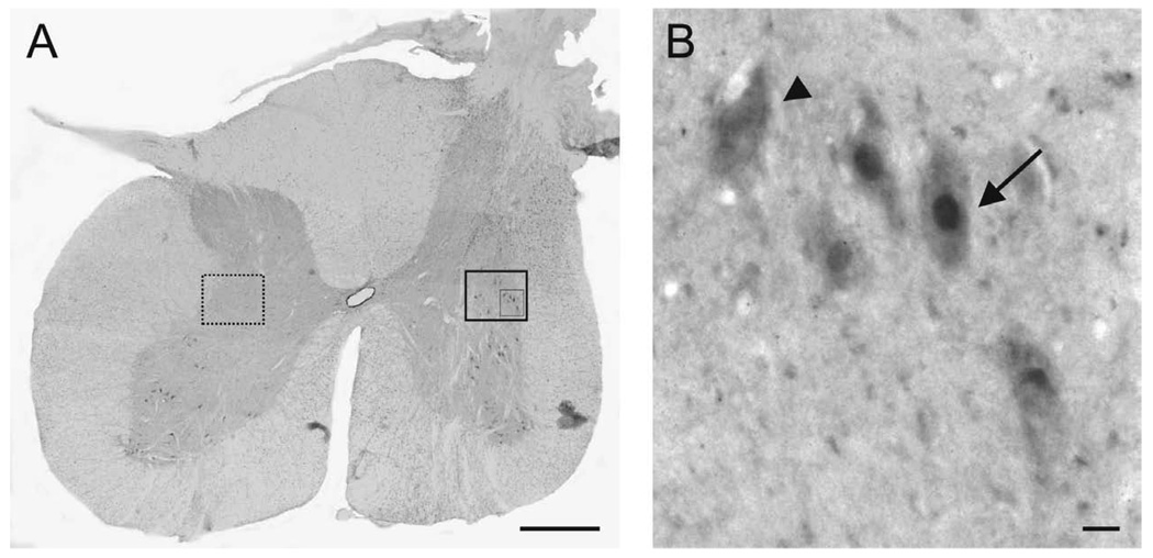
Figure 7.
Induction of c-Fos in host neurons after stimulation of the PNG. (A) Representative transverse section of the spinal cord from a cat at the distal apposition site for the PNG. c-Fos-ir neurons (large boxed area) are located close to the distal apposition site in the intermediate gray, but no c-Fos-ir neurons are present in a comparable area (dashed box) on the contralateral side. Scale bar, 1mm. (B) Higher magnification of the inset boxed area. Arrow: c-Fos-ir neuron. Arrowhead: neuron not expressing c-Fos. Scale bar, 10µm.

