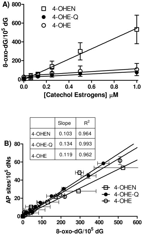
Figure 6. Formation of 8-oxo-dG in calf thymus DNA by catechol estrogens; correlation with AP sites.
A) Concentration-dependent formation of 8-oxo-dG. Calf thymus DNA (100 μg) was treated with catechol/o-quinones in the presence of CuCl2 (10 μM) and NADPH (200 μM). Assays were performed in 10 mM PPB (pH 7.4) at 37 °C for 4 h. DNA was isolated and enzymatically hydrolyzed to deoxynucleosides for LC-MS/MS analysis. B) The regression analysis of 8-oxo-dG versus the number of AP sites. Each point represents the average ± S.D. of three determinations.

