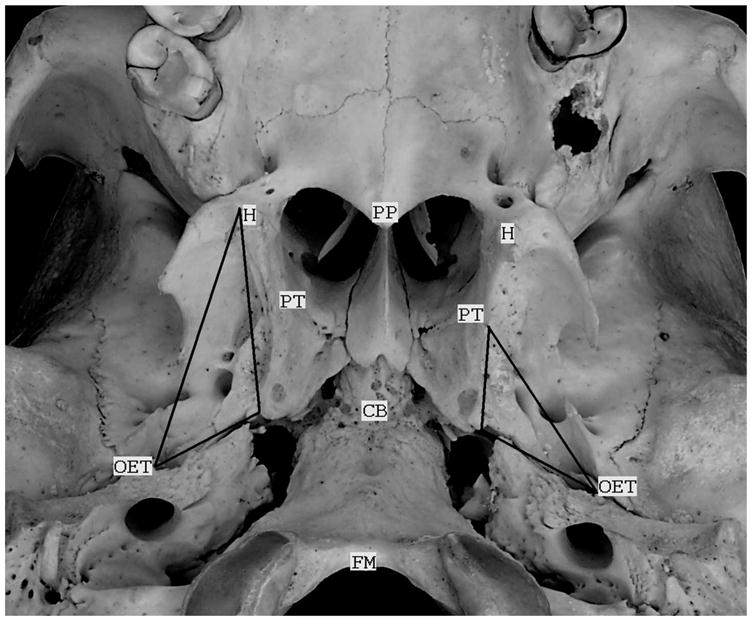
Figure 1.

A photograph of the cranial base of an adult skull with some of the landmarks used for the reconstructions in the present study labeled. These include: the hamular processes (H), the cranial base (CB), the osseous orifices of the ET (OET), the medial pterygoid tubercles (PT), the posterior border of the hard palate (PP) and anterior margin of the foramen magnum (FM). The triangle on the left (right side of the skull) represents the borders of the mTVP muscle and that on the right represents the borders of the mcET. For both, the base of the triangle demarcates the sulcus tubaris. Note that the field of view is tilted to the right and forward in the photograph.
