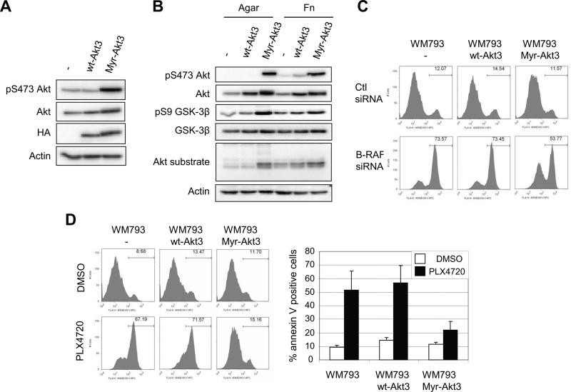
Figure 3.
Expression of constitutively active Akt3 protects melanoma cells from B-RAF inhibition. A, Whole cell lysates from WM793, WM793 HA-Akt3 or WM793 Myr-HA-Akt3 cells were analyzed by Western blotting for HA, total Akt and phosphoS473-Akt. Actin is the loading control. B, WM793, WM793 HA-Akt3 or WM793 Myr-HA-Akt3 cells were serum starved overnight and replated on agar or fibronectin-coated dishes for 1 h in serum-free medium. Cell lysates were analyzed by Western blotting for phospho-S473 Akt, total Akt, phospho GSK3β, GSK3β, and phospho Akt substrate. Actin was used as a loading control. C, WM793, WM793 HA-Akt3 or WM793 Myr-HA-Akt3 cells were transfected with either control or B-RAF siRNAs for 72 h. Cells were then seeded in 3-D collagen gels and cultured in serum-free medium. After 48 h, cells were analyzed by annexin V staining. D, Serum starved WM793, WM793 HA-Akt3 or WM793 Myr-HA-Akt3 cells were seeded in 3-D collagen gels and cultured in serum-free medium containing DMSO or 0.5 μM PLX4720. After 48 h, cells were analyzed as in C. The quantitation of mean −/+ SD from three independent experiments is shown.

