Figure 5.
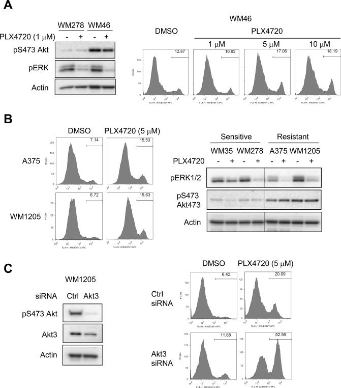
Akt activity in 3-D is elevated in PLX4720-resistant cell lines. A, WM278 and WM46 cells were seeded into 3-D collagen gels −/+ PLX4720 (1, 5 and 10 μM, as indicated). Cells were lysed after 24 hours for Western blot analysis for phosphoS473 Akt, phospho ERK1/2 and actin (left panels). WM46 cells were analyzed after 48 hours for annexin V staining (right panels). B, A375 and 1205Lu cells were seeded into 3-D collagen gels. Left panels show annexin V staining following 48 hours treatment with 5 μM PLX4720. Right panels show Western blot analysis for phosphoS473 Akt, phospho ERK1/2 and actin in A375 and 1205Lu cells in 3-D compared to WM35 and WM278. C, 1205Lu cells were transfected with control non-targeting or Akt3 siRNAs for 72 hours. Cells were lysed for Western blot analysis for phosphoS473 Akt, Akt3 and actin (left panels). Cells were seeded in 3-D collagen gels −/+ 5 uM PLX4720. After 48 hours, cells were harvested for annexinV staining and flow cytometry analysis (right panels).

