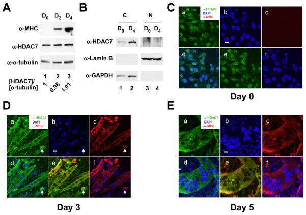
Fig. 1.
Subcellular localization of HDAC7 during myogenesis. (A) The expression levels of HDAC7 during C2C12 differentiation. C2C12 myoblasts were induced to differentiate and cells were harvested at the indicated times. Whole cell extracts were prepared and subjected to immunoblotting with anti-MHC, anti-HDAC7, and anti-α-tubulin antibodies. The relative expression levels of HDAC7 at day 0, 2, or 4 post differentiation are normalized by α-tubulin and are shown. (B) Subcellular fractionation of C2C12 myocytes and differentiated myotubes. Undifferentiated myocytes (D0) and differentiated myotubes (D4) were harvested and subcellular fractions prepared as described in “Materials and Methods”. Cytoplasmic and nuclear fractions of equal cell numbers were loaded followed by SDS-PAGE and Western blotting with anti-HDAC7, anti-Lamin B, or anti-GAPDH antibodies. (C) Undifferentiated C2C12 myoblasts were stained with anti-HDAC7 and anti-MHC antibodies followed by confocal microscopy. Upon induction of differentiation, cells were fixed and stained at days 3 (D) & 5 (E) with DAPI (blue) and antibodies against muscle-specific myosin heavy chain (MHC) (red) followed by confocal microscopy. The white arrow marks a myocyte that expresses MHC with nuclear HDAC7 staining. Scale bars: 20 μm. For C-E, panel a: anti-HDAC7 antibody staining; panel b: DAPI staining; panel c: anti-MHC antibody staining; panel d: merged image of DAPI and anti-HDAC7 antibody staining; panel e: merged image of anti-HDAC7 and anti-MHC antibody staining; panel f: merged image of DAPI and anti-MHC antibody staining.

