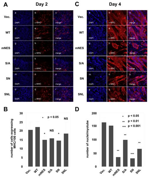
Fig. 3.
The effects of HDAC7 mutations on the ability of HDAC7 to inhibit myogenesis. C2C12 cells stably expressing vector alone, HDAC7 (WT), HDAC7 (S/A), HDAC7 (mNES), HDAC7 (SN), or HDAC7 (SNL) were grown to 80% confluence and induced for differentiation as described in “Materials and Methods”. Cells were fixed and stained at days 2 (A) & 4 (C) with DAPI (blue) and antibodies against muscle-specific MHC (red) followed by epifluorescence microscopy. Representative images obtained from two independent clones of each stable cell line are shown. (B) The average number of cells expressing MHC per 100 cells at day 2. (D) The average number of nuclei per myotubes at day 4. The p values of statistical t-test comparing C2C12 cells expressing wild-type HDAC7 or mutant proteins recorded under the same conditions are shown (* p < 0.05, ** p<0.01, or *** p < 0.001). NS: not significant.

