Fig. 4.
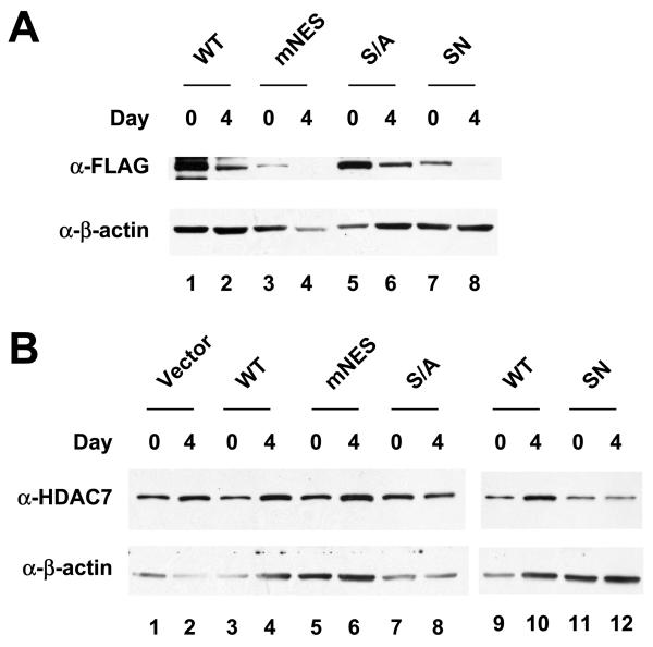
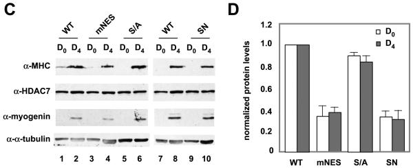
Expression of MHC and myogenin in HDAC7 nuclear mutants. Whole cell extracts of HDAC7 wild-type and mutants were prepared at day 0, 2, and 4. Immunoblotting was performed to detect expression of exogenous FLAG-HDAC7 (A) or total HDAC7 (B). Whole cell extracts of HDAC7 wild-type and mutants were prepared at day 0, 2, and 4. Immunoblotting was performed to detect expression of HDAC7, MHC, and myogenin (C). The relative expression levels of MHC (white column) and myogenin (shaded column) at day 4 (D) were quantified by Photoshop software. The ratio of [MHC]/[a-tubulin] and [myogenin]/[a-tubulin] of HDAC7 (WT) at day 4 was set at 1.


