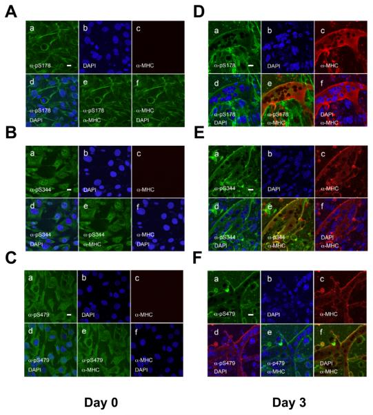
Fig. 7.
Subcellular localization of phosphorylated Class IIa HDACs in myoblasts and differentiated myotubes. Undifferentiated (A-C) and differentiated (D-F) C2C12 cells were stained with anti-pS178, anti-pS344 and anti-pS479 antibodies followed by confocal microscopy. Note that α-pS178 antibodies co-stained with actin filaments and the plasma membrane before and after muscle differentiation. Induction of myoblast differentiation was performed as described in ”Materials and Methods”. Scale bars: 20 μm. panel a: anti-phospho-HDAC7 antibody staining; panel b: DAPI staining; panel c: anti-MHC antibody staining; panel d: merged image of DAPI and anti-phospho-HDAC7 antibody staining; panel e: merged image of anti-phospho-HDAC7 and anti-MHC antibody staining; panel f: merged image of DAPI and anti-MHC antibody staining.

