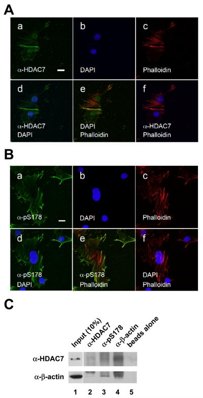
Fig. 8.
pS178-HDAC7 colocalizes with actin filaments. (A) HDAC7 partially colocalizes with actin filaments in C2C12 cells. (B) pS178-HDAC7 colocalizes with actin filaments. Immunostaining was carried out as in Fig. 1 except that the cells were not confluent. Note that DAPI and HDAC7 staining were taken from two different sections. Scale bars: 20 μm. Panel a: anti-HDAC7 antibody staining; panel b: DAPI staining; panel c: anti-MHC antibody staining; panel d: merged image of DAPI and anti-HDAC7 antibody staining; panel e: merged image of anti-HDAC7 and anti-MHC antibody staining; panel f: merged image of DAPI and anti-MHC antibody staining. (C) HDAC7 and actin associate in C2C12 cells. Whole cell lysates prepared from C2C12 myocytes collected at day 0 prior to switching to differentiation medium were subjected to coimmunoprecipitation with beads alone or the indicated antibodies followed by Western blotting with the indicated antibodies.

