Figure 1.
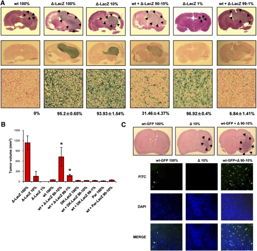
Tumor growth enhancement induced by mixing of wtEGFR and ΔEGFR-expressing cells. (A, top) H&E at day 12 after intracranial injection of U87wt (wt), U87Δ-LacZ (Δ-LacZ), or U87wt mixed with U87Δ-LacZ at 90:10 or 99:1 ratios (wt + Δ-LacZ 90%–10% or wt + Δ-LacZ 99%–1%; 100% = 5 × 10⁵ cells). (Bottom) Whole-brain sections and X-Gal staining of tumor samples; Fast red counterstain. LacZ-positive percentage mean of each tumor sample is indicated below X-Gal staining pictures. (B) Tumor volume after subcutaneous injection of U87wt cells alone or mixed with U87Δ-LacZ, U87Par-LacZ, or U87DK-LacZ at ratios of 90:10 or 99:1 (100% = 1 × 10⁶ cells). Error bars represent mean ± SEM; n = 6. (*) P < 0.05. (C, top) H&E of mouse brains 22 d after intracranial injection of mAstr-Ink4/Arf−/−-wtEGFR-GFP astrocytes (wt-GFP) alone, mAstr-Ink4/Arf−/−-ΔEGFR (Δ) alone, or mAstr-Ink4/Arf−/−-wtEGFR-GFP astrocytes and mAstr-Ink4/Arf−/−-ΔEGFR mixed at a 90:10 ratio (100% = 5 × 10⁵ cells). (Bottom) mAstr-Ink4/Arf−/−-wtEGFR-GFP cells as GFP immunofluorescence. DAPI (blue) labels nuclei.

