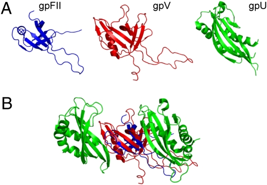
Fig. 5.
Protein structure comparisons with the λ tail terminator protein, gpU. (A) gpFII and gpU were structurally aligned to gpV (rmsds of 2.4 Å over 46 residues and 4.5 Å over 72 residues, respectively) and the structures are displayed side by side in their aligned orientations. (B) gpFII (blue) and gpV (red) are positioned within the gpU hexameric ring structure (half the ring is shown) based on their structural superimposition upon the monomeric structure of gpU. The flanking subunits of gpU are shown (green) with gpFII and gpV in the center. This view shows the inside of the gpU ring from with the same orientation as the rings shown in Fig. 3.

