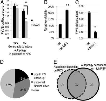
Fig. 1.
Suppression of ROS and expression of Bcl-2 lead to decrease in the levels of autophagy and the type III PI3 kinase activity. (A) Quantification of average type III PI3 kinase activity following knock-down of genes able (yes) and unable (no) to induce autophagy in the presence of NAC. H4 FYVE-dsRed cells were transfected with hit siRNA for 72 h, followed by fixation and imaging on a high-throughput fluorescent microscope at 10× magnification. Average z-scores as compared with nontargeting siRNA are shown. (B) Comparison of the relative average viability of WT or pBabe-Bcl-2–expressing H4 cells transfected with hit gene siRNAs for 72 h. (C) Comparison of average type III PI3 kinase activity in WT or pBabe-Bcl-2–expressing H4 FYVE-dsRed cells following hit siRNA transfection for 72 h. (D) Subdivision of hits whose knock-down was able to induce autophagy under conditions of low PtdIns3P into functional categories based on their ability to up-regulate type III PI3 kinase activity or to alter lysosomal processing. (E) Subdivision of genes whose knock-down led to the induction of autophagy into functional categories based on their dependence on ROS and elevated levels of PtdIns3P (PI3P). *P < 0.05, **P < 0.01 based on two-tailed t test with equal variance. All error bars indicate SEM.

