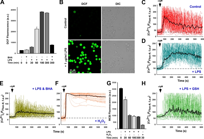
Figure 1.
LPS-induced oxidative stress alters B cell receptor calcium signaling via the depletion of cellular GSH. (A) DT40 cells loaded with ROS indicator H2DCF-DA were challenged with 1 µg/ml LPS for the indicated time period and DCF fluorescence (arbitrary units [au]) measured to assess cellular oxidants either in the absence or presence of 100 µM of the antioxidant compound BHA. Values are representative of three independent experiments. (B) Representative images of DCF fluorescence in DT40 cells in response to 5-h LPS. DIC, differential interference contrast. (C and D) DT40 cells loaded with Fluo-4 were activated by addition of 1.5 µg/ml αIgM and fluorescence recorded via confocal microscopy in control cells (C; n = 33) and after a 5-h 1 µg/ml LPS treatment (D; n = 33). fau, fluorescence arbitrary units. Black traces are mean values of all traces, and baseline fluorescence is indicated as dashed lines. (E) DT40 cells were pretreated with 100 µM BHA before addition of LPS (n = 33). (F) αIgM evoked Ca2+ mobilization in response to direct oxidant challenge with 100 µM H2O2 for 20 min (n = 34). (G) DT40 cells activated with either LPS or H2O2 were loaded with the GSH cross-linking fluorophore MCB to assess reduced GSH levels via the GSH-MCB conjugate. (H) Calcium mobilization after αIgM addition in DT40 cells challenged with LPS and supplemented with 2.5 mM cell-permeable GSH-ester. αIgM addition is noted by arrows. Error bars indicate mean ± SEM.

