Figure 5.
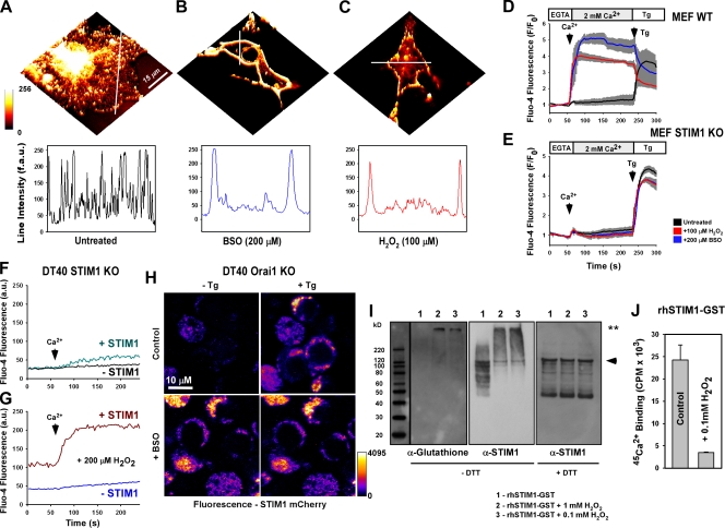
Oxidative stress triggers store-independent STIM1 redistribution and CRAC activation via S-glutathionylation. COS7 cells were transfected with WT STIM1 fused to the mCherry fluorescent construct. 36 h after transfection, STIM1 distribution was visualized via confocal microscopy. (A) STIM1 presents as an ER-resident protein in unstimulated cells, as demonstrated by fluorescence intensity through the perinuclear region. (B and C) STIM1 redistribution to discrete puncta near the plasma membrane is observed after either 200 µM BSO challenge for 24 h (B) or 20-min 100 µM H2O2 treatment (C). (A–C) Line scans indicate the distribution of STIM1-mCherry before and after the treatment. (D and E) Fluo-4–loaded WT MEF cells exposed to either BSO or H2O2 exhibited capacitive calcium entry without ER calcium store depletion by 2 µM Tg (D) that was not observed in STIM1 KO MEFs (E). Traces represent the mean fluorescence of all cells in the microscopic field. Values representing three independent experiments are displayed as a gray bar surrounding each trace. (F and G) Addition of 2 mM Ca2+ to the extracellular buffer in control (F) and after 20-min exposure to 200 µM H2O2 (G) in DT40 STIM1 KO cells. H2O2 challenge elicited store-independent Ca2+ entry upon addition of Ca2+. STIM1-mCherry–negative cells were used as controls (n = 3). (H) Orai1 KO DT40 cells transfected with WT STIM1 mCherry before and 2.5 min after addition of 2 µM Tg. (I) Recombinant human STIM1 protein (rhSTIM1-GST) in buffer containing 10 mM GSH was incubated with 1 mM and 0.1 mM H2O2 for 30 min and resolved under nonreducing conditions. (left) Probed with α-glutathionylation antibody (**, S-glutathionylated STIM1). (middle) Stripped membrane reprobed with α-STIM1 antibody. (right) Probed with α-STIM1 antibody under reducing conditions. The arrowhead indicates native recombinant protein. Black line indicates that intervening lanes have been spliced out. (J) 200 µM 45Ca2+-binding affinity (Luik et al., 2008; Stathopulos et al., 2008) of recombinant human STIM1 after incubation with 100 µM H2O2 for 30 min. 45Ca2+ was assessed by liquid scintillation counting of duplicate protein filtrate samples and background corrected (n = 5). Error bars indicate mean ± SEM.

