Figure 3.
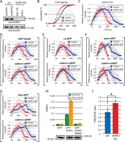
Loss of SHIP2 affects PI(4,5)P2 levels and accelerates endocytic CCP dynamics. (A) Anti-SHIP2 and anti–β-actin Western blots of COS-7 cells stably expressing control shRNA (ctrl) or SHIP2-specific shRNA (SHIP2 KD). (B) Cumulative histograms of CCP lifetime (GFP–clathrin light chain signal) in control cells (n = 697) and SHIP2 KD cells (n = 1,648). (C) Time course of relative fluorescence intensity of GFP-clathrin in control cells and SHIP2 KD cells. (D–G) Time course of relative fluorescence intensity of mRFP and GFP fluorescence in cells double transfected with clathrin-mRFP and with GFP fusions of clathrin adaptors and dynamin as indicated. (H, top) PI(4,5)P2 and PI(3,4,5)P3 phosphatase activity (Malachite green assay) in anti-GFP immunoprecipitates from COS-7 cells expressing GFP, GFP-SHIP2, or GFP–SHIP2-D608A (catalytically dead). (bottom) Anti-GFP Western blot of the material used in the phosphatase assay. (I) PIP2/PIP ratio in control and SHIP2 KD cells as determined by HPLC (*, P < 0.05; t test). (C–I) Error bars show mean ± SD.

