Figure 6.
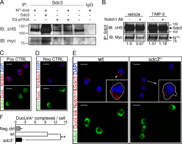
Syndecan-3 and Notch are in a complex on the SC membrane. (A) Syndecan-3 and Notch1 (NFL-6mt) were coimmunoprecipitated from MM14 cell extracts with an anti–Syndecan-3 antibody 24 h after transfection, and Western blots were probed for the myc-tag epitope and for Syndecan-3 using a terminal heparan sulfate attachment antibody (Δ-HS). (B) Extracts from MM14 cells cultured in the presence or absence of an anti-Notch1 antibody, transfected as in E, and treated with or without 200 nM TIMP-3 were immunoprecipitated with an anti–Syndecan-3 antibody. Blots probed for the myc-tag epitope, then stripped and reprobed for Δ-HS. The relative levels of NTM were quantified and plotted with the level of NTM in the untreated sample set to 1. Molecular mass standards are indicated next to gel blots (kD). (C–F) Endogenous Notch and Syndecan-3 are present in a protein complex on the SC surface, as revealed using DuoLink with an extracellular anti-Notch1 antibody and an intracellular Syndecan-3 antibody. Two distinct Notch1 antibodies from different species show as positive complexes in Syndecan-4 (sdc4)+ cells (green) after DuoLink labeling (C, red), whereas elimination of the primary anti-Notch1 and anti–Syndecan-3 antibodies yielded no positive complexes (D). Endogenous Notch1 and Syndecan-3 appear as complexes (red dots) in wild-type Syndecan-4+ cells (E) but are absent or at background levels in sdc3−/− cells (E). Arrows indicate which cells are magnified in the inset panels. (F) Quantification of C–E, where the mean number of DuoLink+ complexes/cell is plotted. n = 3. Open bars, wild type; shaded bars, sdc3−/−. Error bars indicate SEM. **, P < 0.01; #, P > 0.05. Bars, 10 µm.

