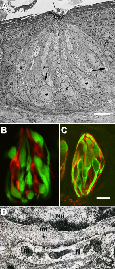
Figure 2.
Cell types and synapses in the taste bud. (A) Electron micrograph of a rabbit taste bud showing cells with dark or light cytoplasm, and nerve profiles (arrows). Asterisks mark Type II (receptor) cells. Reprinted with permission from J. Comp. Neurol. (Royer and Kinnamon, 1991). (B) A taste bud from a transgenic mouse expressing GFP only in receptor (Type II) cells. Presynaptic cells are immunostained (red) for aromatic amino acid decarboxylase (a neurotransmitter-synthesizing enzyme that is a marker for these cells), and are distinct from receptor cells, identified by GFP (green). Reprinted with permission from J. Neurosci. (C) Taste buds immunostained for NTPDase2 (an ectonucleotidase associated with the plasma membrane of Type I cells) reveal the thin lamellae (red) of Type I cells. These cytoplasmic extensions wrap around other cells in the taste bud. GFP (green) indicates receptor cells as in B. Bar, 10 µm. Image courtesy of M.S. Sinclair and N. Chaudhari. (D) High magnification electron micrograph of a synapse between a presynaptic taste cell and a nerve terminal (N) in a hamster taste bud. The nucleus (Nu) of the presynaptic cell is at the top, and neurotransmitter vesicles cluster near the synapse(s). The nerve profile includes mitochondria (m) and electron-dense postsynaptic densities. mt, microtubule. Image courtesy of J.C. Kinnamon.

