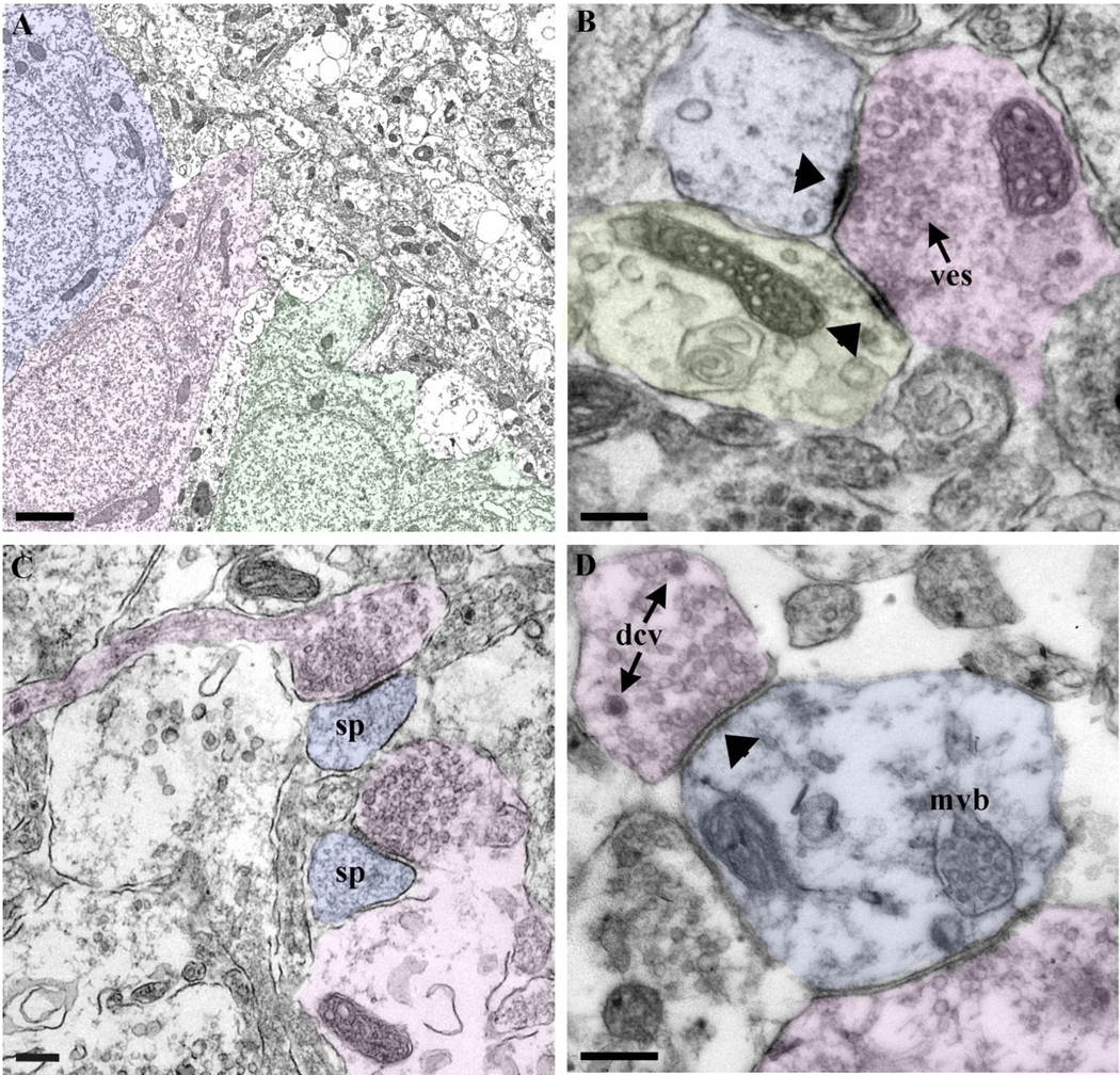
Figure 3.
Ultrastructural organization of the stage 45 Xenopus optic tectum. A: Low-magnification electron micrograph of the medial portion of the optic tectum shows three neuronal cell bodies positioned adjacent to the tectal neuropil. B: Fully mature synapses are established between a presynaptic axon terminal (highlighted in pink) and two postsynaptic dendrites (blue and yellow). Arrowheads point to the postsynaptic densities (ves; synaptic vesicles). C: Presynaptic terminals (pink) also establish mature synaptic contacts with dendritic spines (blue). D: The presence of dense-core vesicles presynaptically (dcv) and multivesicular bodies (mvb) in postsynaptic terminals (blue) in the developing tectal neuropil is also shown in this sample tissue embedded in LR-white. Scale bars = 2µm in A; = 200 nm in B – D.

