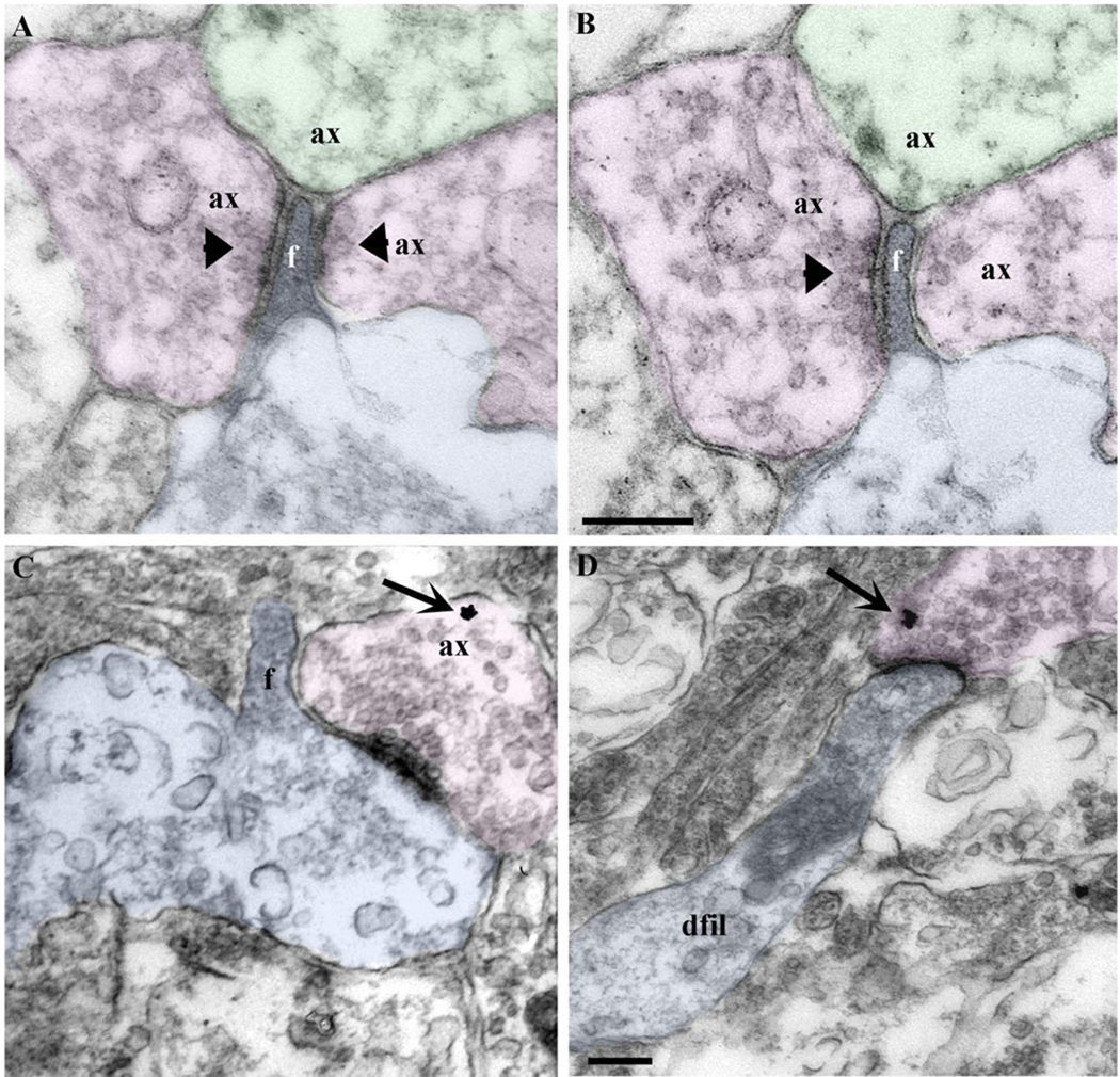
Figure 5.
Filopodial synapses in the stage 45 Xenopus optic tectum. A,B: Serial ultrathin sections show a nascent dendritic filopodium (blue) that receives synaptic input (arrowheads) from multiple presynaptic terminals (pink). A nonsynaptic terminal containing a few synaptic vesicles (green) also contacts this filopodium. C,D: RGC axon terminals (pink) labeled with YFP and identified by gold particle immunoreactivity (arrows) establish synaptic contacts with a filopodia-bearing (f) growth cone-like structure (blue) in C, and with dendritic filopodial-like profiles (dfil; blue) in D. Dendritic filopodia-like profiles can receive multiple synaptic contacts (A) or can receive a single synaptic input from a presynaptic fiber (D). Scale bars = 200 nm in B (applies to A,B); 200 nm in D (applies to C,D).

