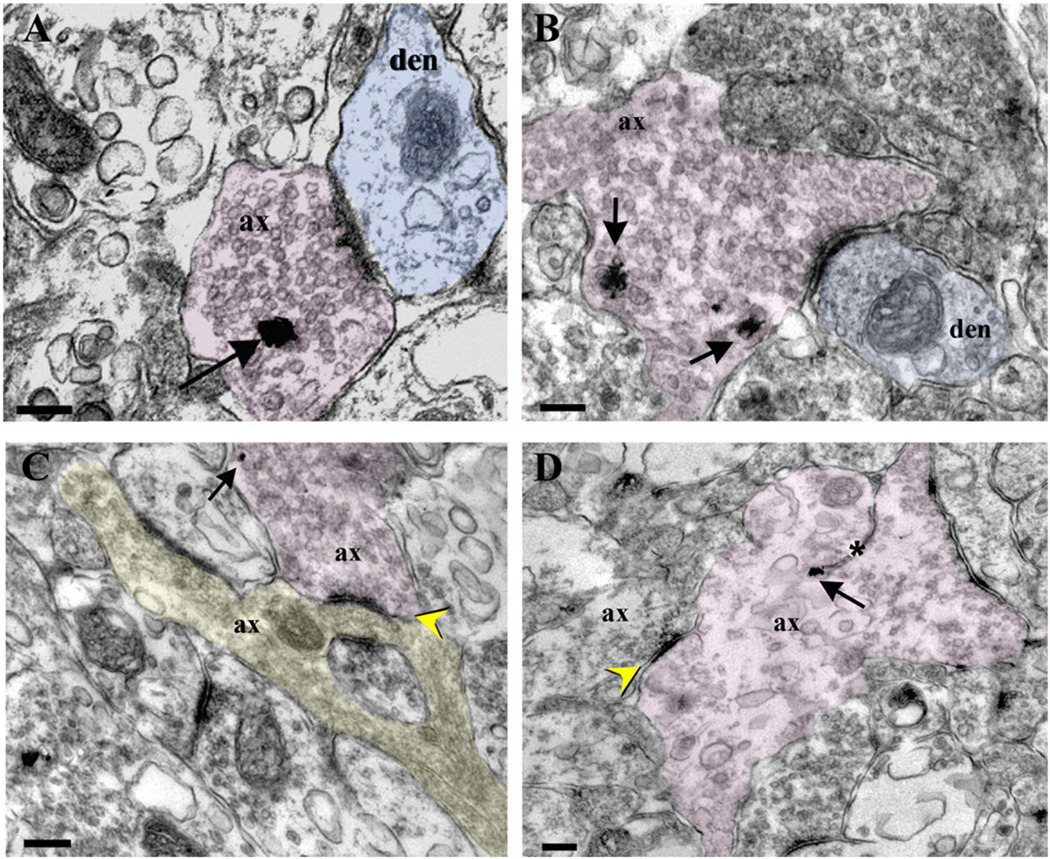
Figure 6.
Shaft and axoaxonal synapses in the Xenopus optic tectum. A,B: Synapses between YFP-immunopositive RGC axon terminals (pink) and tectal neuron dendrites at a shaft (blue) are illustrated in these sections of the stage 45 tectal neuropil. C,D: Axoaxonal synaptic profiles are also found in the stage 45 Xenopus tectal neuropil. C: An RGC axon (pink) expressing YFP (arrow) makes synaptic contact with an axonal terminal (highlighted in yellow). D: In this sample, a YFP-identified RGC axonal profile (pink) receives axoaxonal input from a neighboring axon (yellow arrowhead). A spinule, a thin projection of cytoplasm and membrane of the dendritic surface that divide a presynaptic bouton (Sorra et al., 1998), is marked by the asterisk. In all panels, the silver-enhanced YFP immunogold particles are marked by arrows. ax, Axon terminal; den, dendritic profile. Scale bars = 200 nm.

