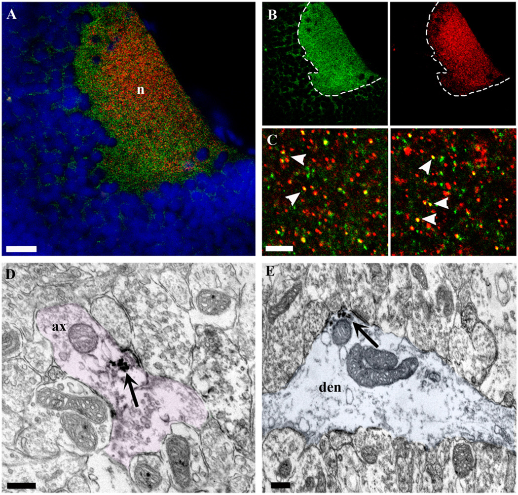
Figure 8.
Cellular and subcellular distribution of full-length TrkB in the stage 45 Xenopus optic tectum. A,B: Coronal section of a stage 45 Xenopus midbrain at the level of the optic tectum shows the distribution of TrkB immunoreactivity (green fluorescence) in the tectal neuropil (n) and its colocalization with the presynaptic marker SNAP-25 (red fluorescence). In A, the cell body layer is shown by DAPI fluorescence (blue). B: The distribution of TrkB immunofluorescence in the neuropil and surrounding cell bodies near the neuropil is better illustrated by separating the two channels, in which TrkB alone (green) and SNAP-25 (red) localization are shown. The border between the cell body layer and the tectal neuropil is demarcated by the dashed line. C: Thin, horizontal, high-magnification confocal sections show the colocalization of TrkB (green) and SNAP-25 (red) immunofluorescence at the level of the tectal neuropil. D,E: Immunoelectron microscopy illustrates the subcellular distribution of TrkB in the tectal neuropil. The immunoperoxidase reaction product (arrows) reveals that TrkB immunoreactivity localizes to axon terminals (D, pink) as well as dendritic profiles (E, blue). In E, a large-caliber dendrite that receives multiple synaptic contacts contains TrkB-immunoreactive precipitate near the postsynaptic membrane (arrow). For a green-magenta version of parts of this figure see Supporting Information Figure 4. Scale bars = 50 µm in A; 5 µm in C; 200 nm in D,E.

