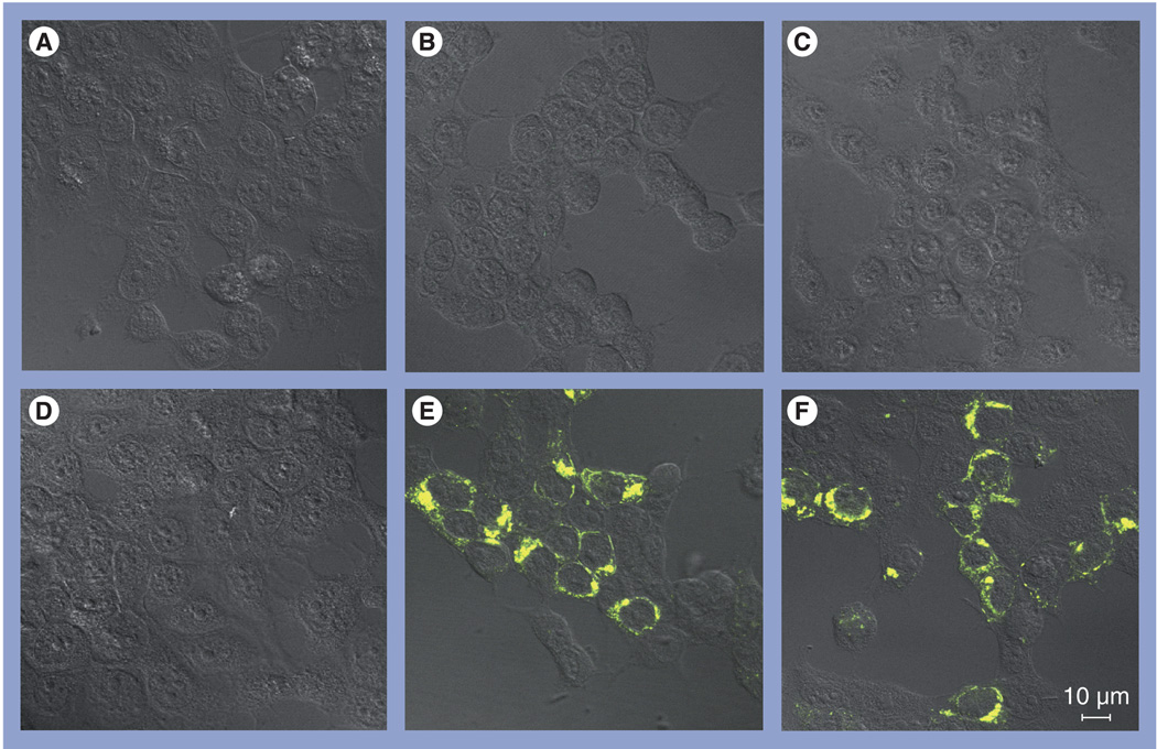
Figure 1. Bimolecular complementation assay.
Human 293T cells were transfected with (A) N-terminal yellow fluorescent protein (NYFP)-tumor susceptibility gene (Tsg)101 alone, (B) C-terminal YFP (CYFP)-Ebola virus (EBOV) VP40 alone, (C) CYFP-Marburg virus VP40 alone, (D) NYFP-Tsg101 plus CYFP-EBOV VP40-ΔPT/PY, (E) NYFP-Tsg101 plus CYFP-EBOV VP40 or (F) NYFP-Tsg101 plus CYFP-Marburg virus VP40. Cells were examined at 24 h post-transfection for YFP fluorescence activity by confocal microscopy using a Zeiss LSM-510 Meta system.

