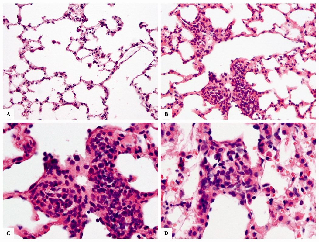
Figure 8.
Micrographs of representative H&E stained lung sections from mice exposed to 124.5 µg/m3 (79.0 µg Ni/m3) of nano-Ni(OH)2 for 5 m. (A) normal lung at 20×; (B) exposed mouse lung section showing chronic inflammatory infiltrate in a terminal duct at 20×; (C) same as previous, showing a predominance of lymphocytes at 40×; (D) inflammatory infiltrate in the pulmonary interstitial space of exposed mouse at 40×.

