
Figure 2.
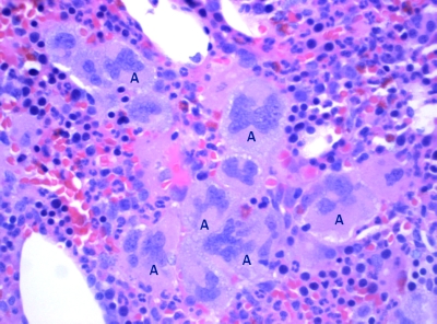
. Histology of trephine bone marrow biopsy (H&E stained) showing large clusters of megakaryocytes (A) with considerable variation in cell size and lobulation of the nucleus. Active erythropoiesis and myelopoiesis showing normal differentiation.