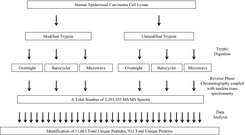
FIGURE 1.
Workflow. This flowchart illustrates the entire sample analysis workflow from start to finish. A human epidermoid carcinoma cell lysate (10 μg) was digested using various in-gel digestion procedures and technologies. The resulting peptides were then analyzed using reverse-phase chromatography coupled with MS/MS. All resulting spectra were then searched against a human protein sequences database using the X! Tandem algorithm for peptide identification.

