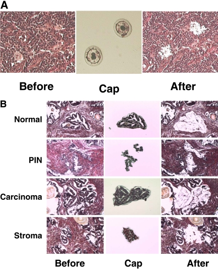
FIGURE 3.
Capture of pure cell populations by LCM. (A) LCM of HL and RS cells. A HL tissue section was stained with H&E (left), and the giant RS cells were captured successfully by LCM and visualized on the LCM cap (middle). (B) LCM of pure cell populations from a prostate cancer sample. A prostate adenocarcinoma tissue section was stained with H&E (left), and the benign, prostatic intraepithelial neoplasia (PIN), malignant, and stromal cells were isolated by LCM, respectively, and visualized on the LCM cap (middle). All human tissues were obtained from patients who provided informed consent and acquired through the Hollings Cancer Center Tissue Biorepository in accordance with an Institutional Review Board-approved protocol.

