
Figure 4. MRI-based fetal phenotypic analysis.
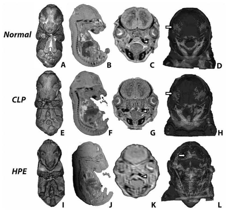
3-D volume (A,E,I) and surface rendering (D,H,L) and 2D sections (B,C,F,G,J,K) reveal external and internal CLP- and HPE-associated dysmorphology. Cleft lip is indicated by black arrows while white arrowheads note secondary palate structure. White arrows note cranial frontal bones which are abnormally fused (metopic craniosyntosis) in the HPE fetus (L).