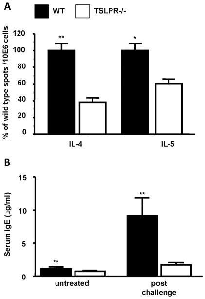
Figure 3. Reduced features of a Th2 allergic response in FITC sensitized TSLPR−/− mice.
(a) Cytokine secretion by PMA/ionomycin stimulated skin-draining lymph node cells one week following sensitization of WT and TSLPR−/− mice with 0.5% FITC in acetone/dibutyl phthalate. The results are expressed as percent of WT cytokine forming spots/106 cells from three independent experiments (**P < 0.01, Wilcoxon test, n=15). (b) Serum IgE levels in untreated or FITC sensitized and challenged WT or TSLPR−/− mice. WT mice have significantly higher basal and post-challenge serum IgE levels (**P < 0.01, Wilcoxon test, n≥9 for each group).

