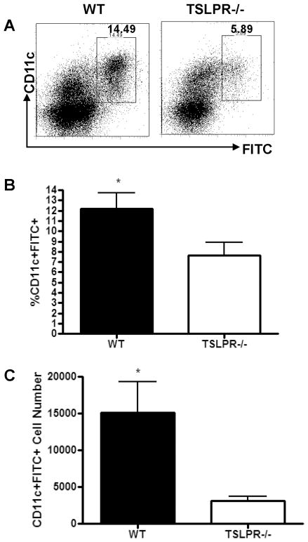
Figure 4. Reduced accumulation of FITC+CD11c+ cells in skin draining lymph nodes of TSLPR−/− mice 24 H post-sensitization with FITC in acetone/DBP.
(a) Representative FACS plots from skin-draining inguinal and axillary lymph node cells from WT and TSLPR−/− mice 24 H post-sensitization on the abdomen with 0.5% FITC, after Percoll enrichment for low density cells. Values represent frequency of FITC+CD11c+ cells within the live cell gate. (b) Frequency and (c) absolute number of FITC+CD11c+ cells found in inguinal and axillary LN of WT and TSLPR−/− mice 24 H post FITC sensitization. TSLPR−/− FITC+CD11c+ cell frequency and number are significantly reduced compared to WT. (*P<0.05, Students T-test, results pooled from two independent experiments, n=7 mice per group).

