
Figure 1.
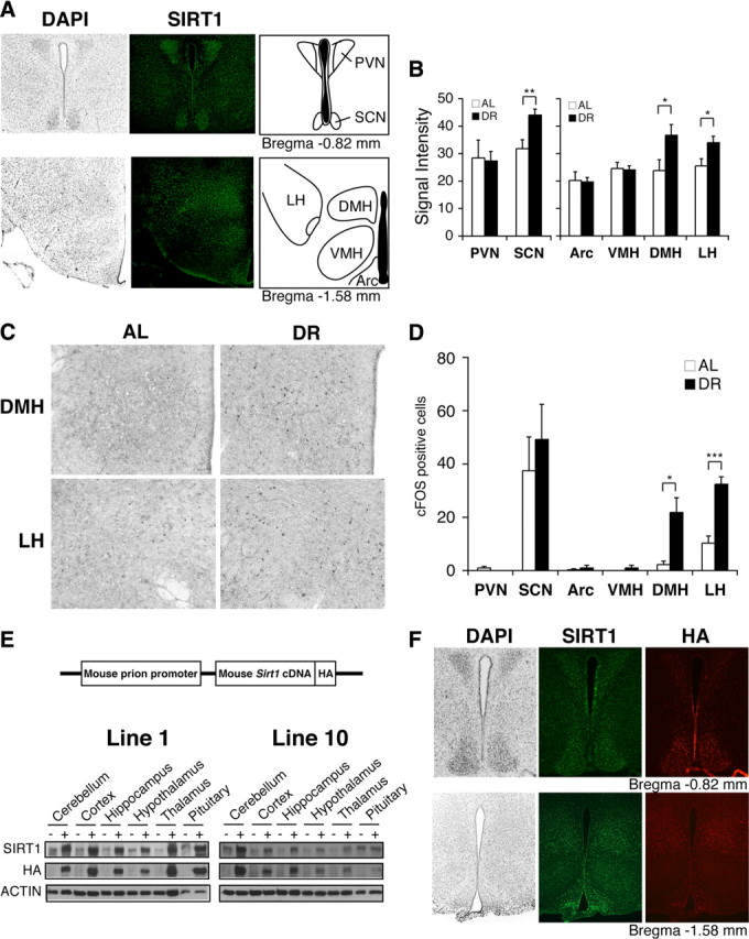
SIRT1 is expressed in major hypothalamic nuclei, and both SIRT1 levels and the number of activated neurons increase in the DMH and LH in diet-restricted hypothalami. A, Immunofluorescent staining of SIRT1 (green) in major hypothalamic nuclei, including the PVN, SCN, Arc, VMH, DMH, and LH. Nuclei are counterstained by DAPI (gray). B, Signal intensities of SIRT1 staining in hypothalami after 14 d DR compared to AL (*p < 0.05, **p < 0.01, n = 4 mice, 4–8 sections per hypothalamic nucleus). The signal intensity per area was digitally quantified after subtracting surrounding background. Results are shown as mean values ± SEM. C, D, cFOS staining in the DMH and LH after 14 d DR (C), and quantification of the number of cFOS-positive cells in hypothalamic nuclei (*p < 0.05, ***p < 0.001, n = 4 mice, 4–8 sections per hypothalamic nucleus). The numbers of cFOS-positive cells are shown as mean values ± SEM (D). E, Transgene structure for the production of BRASTO mice (upper panel) and distribution of SIRT1 in major brain regions in both lines 1 and 10 of BRASTO mice (lower panels). F, Immunofluorescent staining of SIRT1 (green) and HA (red) in hypothalami of BRASTO line 10. Nuclei are counterstained by DAPI (gray).