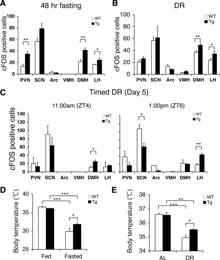
Figure 3.
BRASTO mice show enhanced neural activation in the DMH and LH in response to multiple diet-restricting paradigms. A–C, Quantification of cFOS-positive cells in major hypothalamic nuclei after 48 h fasting (A), 14 d DR (B), and 5 d of timed DR (C) in BRASTO mice in line 10 (*p < 0.05, **p < 0.01, n = 4 for 48 h fasting and DR, 4–8 sections per hypothalamic nucleus; n = 2 at each time point for timed DR, 2–5 sections per hypothalamic nucleus). The numbers of cFOS-positive cells are shown as mean values ± SEM. D, E, Rectal body temperature of BRASTO mice after 48 h fasting (D) and during 14 d DR (E). Levels of rectal body temperature are shown as mean values ± SEM (*p < 0.05, **p < 0.01, ***p < 0.001 by one-way ANOVA with Tukey–Kramer post hoc test, n = 6–7 for 48 h fasting, n = 6 for DR).

